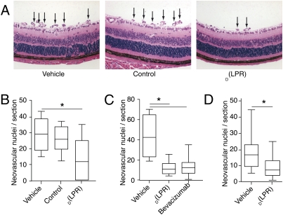
Fig. 4.
Systemic and topical treatment with D(LPR) inhibits retinal angiogenesis in a mouse retinopathy-of-prematurity model. Retinal neovascularization was induced in C57BL/6 neonatal mice by exposure to 75% oxygen (P7–P12) followed by daily i.p. administration of D(LPR) or control (20 mg/kg per day). (A) H&E-stained retinal sections (P19) showed new blood vessels at the retinal inner surface (arrows) in control animals (Top and Center). There was marked reduction in D(LPR)-treated mice (Bottom). (B) P19 quantification of neovascular nuclei protruding into the vitreous space. Serial sections (n > 5) of eyes (n > 10) were quantified in each group. Treatment with D(LPR) yielded a significant reduction in nuclei relative to vehicle or control. (C) In systemic (i.p.) delivery, the magnitude of D(LPR)-induced neovascularization inhibition was similar to that observed by treatment with the monoclonal antibody bevacizumab. (D) Topical delivery of D(LPR) in an eye-drop formulation (200 μg e.d. three times daily) also induced a significant reduction in abnormal retinal angiogenesis. Shown is mean ± SEM in each experiment.*, Student’s t test (P < 0.05).

