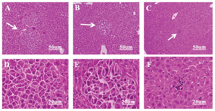
Figure 5.

Histopathology of liver metastases arising from CXCR4 siRNA-transfected OCM3 uveal melanoma cells. OCM3 uveal melanoma cells were transfected with either CXCR4 siRNA or control siRNA. OCM3 uveal melanoma cells (2 × 10⁵) were injected into the spleen capsules of NOD-SCID mice. Mice underwent necropsy 35 days later, and livers were processed for conventional hematoxylin and eosin staining. (A, D) Untreated. (B, E) siRNA control-treated OCM3 uveal melanoma cells. (C, F) CXCR4 siRNA-treated OCM3 melanoma cells. Arrow: metastatic tumor nodules. (A–C) Low power and (D–F) high-power photographs of A–C, respectively.
