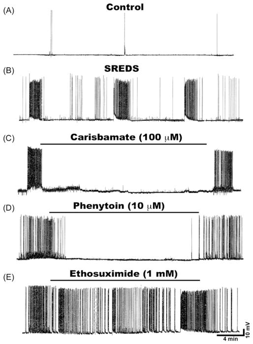
Figure 2.
Effects of various AEDs on SREDs in cultured hippocampal neurons. (A) Current clamp recording from a representative control neuron displaying baseline activity consisting of intermittent action potentials. (B) Recording from an “epileptic” neuron 1-day following a 3-h exposure to low Mg2+ solution demonstrates occurrence of characteristic SREDs. Three SREDs or spontaneous seizure episodes lasting ~60–90 s can be seen in this recording frame. These SREDs occurred throughout the life of the cultures and are indicative of the pathophysiological “epileptic” phenotype. Effects of various AEDs on SREDs in cultured hippocampal neurons. Application of (C) Carisbamate (100 μM) or (D) Phenytoin (10 μM) to the epileptic neurons abolished the expression of SRED activity. However application of (E) Ethosuximide (1 mM) failed to stop SRED activity. The effects of AEDs on SREDs were reversible and seizures reappeared following drug washout.

