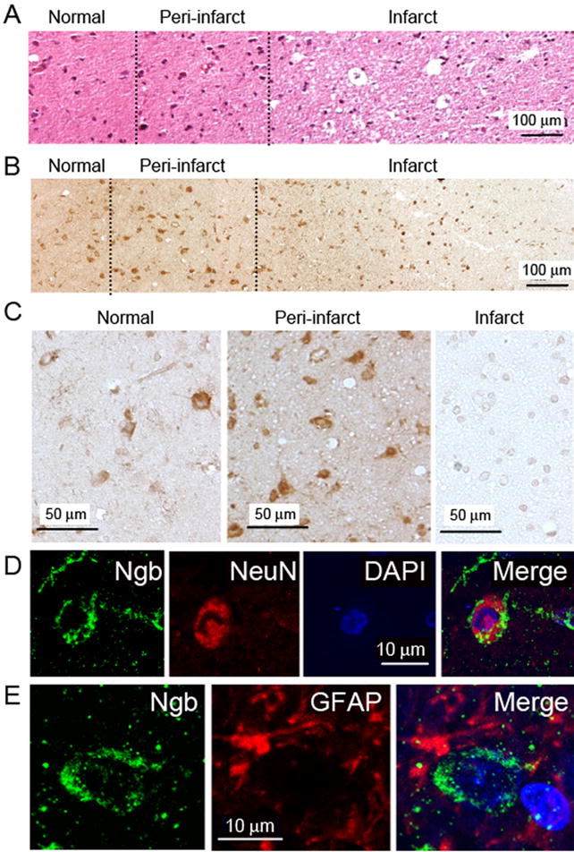
Figure 2. Ngb expression in post-stroke human brain.

(A) Hematoxylin- and eosin-stained section from cerebral cortex of infarcted brain. (B) Ngb immunohistochemistry in cerebral cortex of infarcted brain. (C) Ngb immunohistochemistry in cerebral cortex of infarcted brain at higher magnification. (D) Double-label immunohistochemistry in cerebral cortex of infarcted brain showing expression of Ngb (left, green) and NeuN (center, red), DAPI-stained nuclei (center right, blue) and merged image (right). (D) Double-label immunohistochemistry in cerebral cortex of infarcted brain showing expression of Ngb (left, green) and GFAP (center, red), and merged image (right). Images shown are representative of similar findings in 6/6 patients studied.
