
Figure 3.
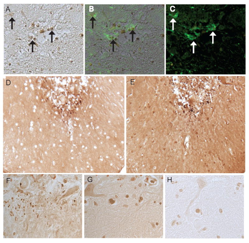
In a representative MCI case, many of the iron-positive structures (A) are associated with glial cells recognized by fluorescence microscopy using GFAP as the antibody (C). B is a merged image showing the iron-stained bodies associated with GFAP-positive glia (arrows). The same structures on adjacent serial sections are labeled using both the detection method with potassium ferrocyanide (D) as well as incubation with only DAB/ H2O2 to detect sites of metal-catalyzed redox activity (E). Redox metal positive structures within the cerebellum of an MCI case (F) are sensitive to chelation treatment with EDTA (G) and deferroxamine (H).