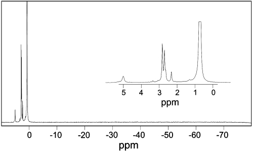
Fig. 1.
A NMR spectrum of the crystallization components, including β-PGM, G6P, and . The spectrum shows (expanded in inset) resonances of the protein-bound phosphate from G6P in the complex (5.00 ppm), free G6P in solution as α- and β-anomers (2.70 and 2.82 ppm, respectively) and free Pi (0.72 ppm), as well as minor amounts of several other non-protein-bound sugar phosphates. No other peaks are observed at chemical shifts resonating upfield of Pi characteristic of a protein-bound aspartyl phosphate or a pentacoordinate phosphorane species.

