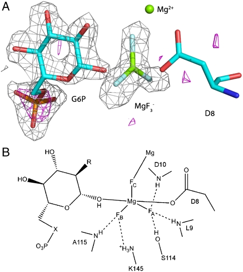
Fig. 2.
Structure of the complex active site. (A) The difference Fourier map and the anomalous substructure of the complex. Anomalous difference density contoured at 3σ is shown as a magenta mesh. A large peak (height ) is visible for the phosphorus atom in G6P. No corresponding phosphorus peak was observed in the active site confirming the assignment of the trigonal planar species as and not . The difference electron density () from the same data is shown as a gray mesh contoured at 3σ for G6P and the moiety before their inclusion in the model. (B) Schematic view of the complex active site. Three sugar moieties were studied: G6P (, ); 6-deoxy-6-(phosphonomethyl)-D-glucopyranoside (, ); 2-deoxy-G6P (, ).

