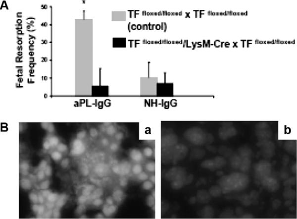
Figure 3.
TF expression by myeloids cells but not fetal-derived cells contributes to aPL-induced decidual oxidative stress and fetal loss. (A) Treatment with aPL-IgG caused an increase in fetal resorptions in TFfloxed/floxed mice (*P < 0.001 versus NH-IgG). TFfloxed/floxed/LysM-Cre mice were protected from fetal loss induced by aPL-IgG. Fetal resorption frequency in these mice was comparable to TFfloxed/floxed mice treated with NH-IgG. (B) Superoxide (O2−) generation in decidual tissue was determined using dihydroethydium fluorescence. aPL-induced O2− formation (a) is attenuated in TFfloxed/floxed/LysM-Cre mice (b) to a similar extent to NH-IgG-treated mice. Original magnification ×800 (originally published by Redecha, et al.45).

