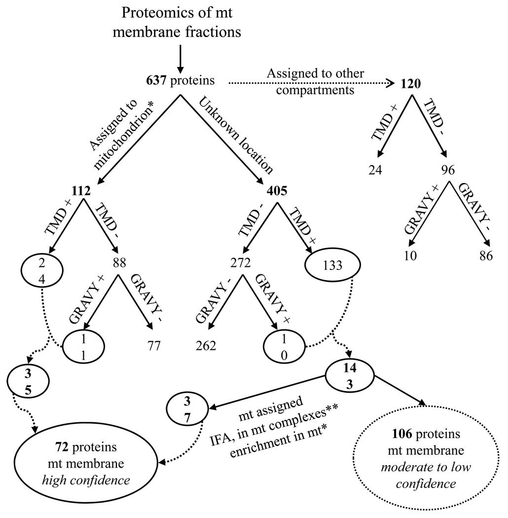
Figure 3.
Grouping of proteins identified from the MS/MS analyses. The number of proteins designated to mitochondria, to another cellular compartment or to an unknown location is indicated. The putative membrane assignment was done by presence of predicted TMD and/or positive GRAVY score. The circled groups were assigned to mt membranes with high confidence, whereas the dotted-circled groups with moderate to low confidence.
* [3]

