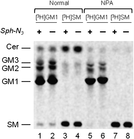
Fig. 4.
Radioactive lipids analysis of normal (lanes 1, 2, 3, 4) or NPA (lanes 5, 6, 7, 8) cells treated (lanes 1, 3, 5, 7) or not (lanes 2, 4, 6, 8) with Sph-N3 and fed with [3-3H(Sph)]GM1(lanes 1, 2, 5, 6) or [3-3H(Sph)]SM (lanes 3, 4, 7, 8). One thousand dpm of the total lipids extracted from normal and NPA fibroblasts fed with Sph-N3 followed by radioactive GM1 or SM administration were separated by HPTLC using CHCl3/CH3OH/(CH3)2CHOH/0.2% aqueous CaCl2, 20:60:20:4 by vol. as solvent system. Radioactive lipids were detected by digital autoradiography performed with a Biospace β-imager instrument for 48 h.

