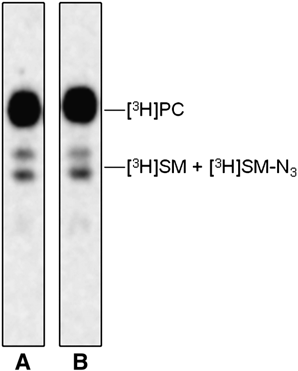
Fig. 5.
Radioactive lipids analysis. TLC separation of the total lipids extracted from normal (lane A) and NPA (lane B) fibroblasts fed with Sph-N3 and [methyl-3H]choline. One thousand dpm were applied on a 4 mm line for each sample. TLC was run in CHCl3/CH3OH/0.2% aqueous CaCl2, 50:40:8 by vol. Digital autoradiography was performed with a Biospace β-imager instrument for 48 h.

