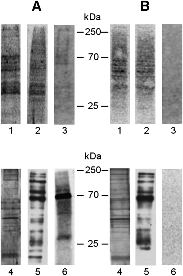
Fig. 9.
Proteins cross-linked with [3H]SM-N3 from cell homogenates (lane 1), HD fraction (lane 2) and DRM fraction (lane 3) prepared from normal (A) and NPA (B) fibroblasts were separated by 10% SDS-PAGE, blotted on a polyvinyldifluoride (PVDF) membrane, and visualized by digital autoradiography for 120 h. Proteins recovered in the immunoprecipitation experiments performed in domain preserving conditions starting from aliquots of DRM fractions prepared from normal (A) and NPA (B) fibroblasts were separated by 10% SDS-PAGE. The proteins were directly detected by silver staining (lane 4) or blotted on a PVDF membrane and then visualized by Western blot using HRP-streptavidin (lane 5) or by digital autoradiography for 120 h (lane 6). Data are the means of three different experiments.

