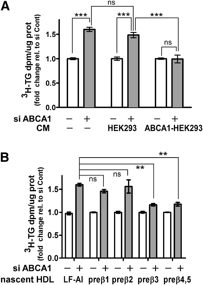
Fig. 6.
Large nascent HDLs attenuate the increased TG secretion induced by ABCA1 silencing. A: Conditioned medium was prepared by incubating 10 μg apoA-I /ml with control and ABCA1-expressing HEK 293 cells for 24 h. Nonconditioned medium (−CM) was prepared by incubating 10 µg/ml of apoA-I with empty dishes for 24 h. The conditioned or nonconditioned medium along with [3H]oleate + 0.4 mM oleate was then added to McA cells that had been previously transfected with control or ABCA1 siRNA (25 nM for 48 h). After additional 12 h incubation, [3H]TG secretion into the medium was quantified as described in Fig. 1 legend. B: Conditioned medium from ABCA1-expressing HEK293 cells was prepared as described in A and fractionated by high resolution FPLC into individual subfractions of nascent HDL as described previously (8). McA cells that had been previously transfected with control or ABCA1 siRNA (25 nM for 48 h) were incubated with individual nascent HDL subfractions (2 µg protein/ml) in the presence of [3H]oleate + 0.8 mM oleate for an additional 12 h and the amount of newly synthesized TG secreted into the medium was quantified. Results are normalized to control siRNA transfected cells and are expressed as mean ± SEM of triplicate analyses. ** (P < 0.01), *** (P < 0.001); ns, not significant, by one-way ANOVA (Tukey's multiple comparison test).

