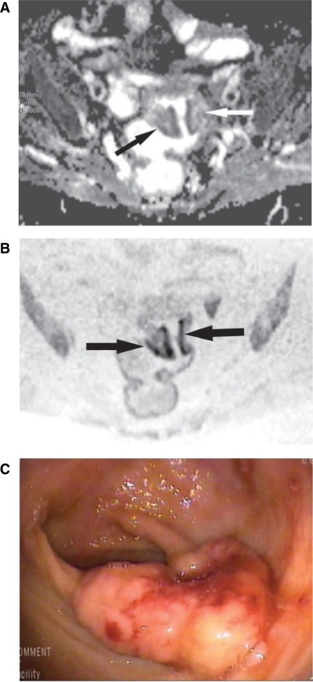
Figure 2.
An 80-year-old woman with adenocarcinoma of the upper third of the rectum, at the rectosigmoid junction. (A) Axial ADC map obtained with b = 1000 s/mm2 shows tumour (arrows). ADC is 1.195 × 10⁻³ mm2/s. (B) Axial FBDW-SSEPI image obtained with b = 1000 s/mm2 and displayed using black and white reversed contrast shows dark well-defined areas (arrows) that were correctly classified as rectal cancer by the 2 reviewers (true-positive case). (C) Optical colonoscopic view confirms the rectal tumour that was classified as a T2 well-differentiated adenocarcinoma at histopathologic analysis after surgical resection.

