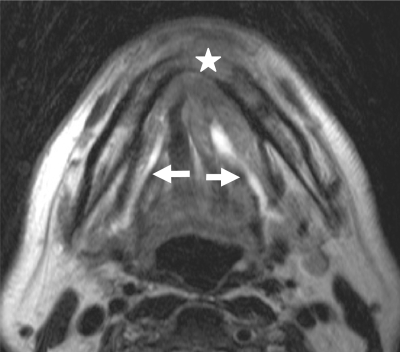
Figure 5.
Axial T2-weighted MR image of a patient with histologically proven squamous cell cancer of the floor of the mouth showing the tumour with involvement of the mandible and extension into the adjacent soft tissues anteriorly (star). Note obstruction with dilatation of the Wharton duct on both sides (arrows).

