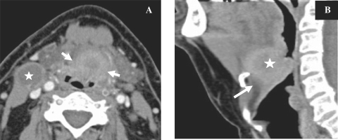
Figure 9.
Axial CT image (A) in a patient with proven SCC of the base of the tongue showing the tumour (arrows) with central necrosis and a necrotic lymph node on the right side (star). The tumour extends into the valleculae on both sides and displaces the epiglottis posteriorly. Sagittal reconstruction (B) showing the tumour (star) with infiltration of the preepiglottic space (arrow).

