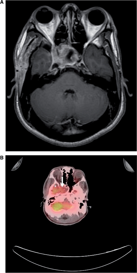
Figure 1.
(A) T1-weighted MR imaging (spin echo, repetition time (TR)/echo time (TE)=466.7 ms/13 ms) with contrast enhancement of the head shows an enhancing mass in the sella turcica extending to the right cavernous sinus, consistent with local recurrence of tumor. (B) Whole-body FDG-PET/CT fusion image in the same level shows abnormal increased FDG uptake of tumor (large arrow) and physiological uptake of right temporal lobe (small arrow).

