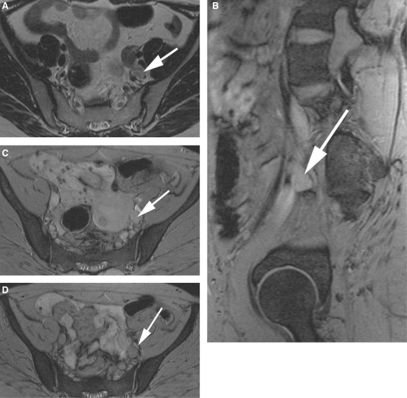
Figure 3.
A 55-year-old patient with endometrial carcinoma. A left interiliac node is seen on the axial T2W image (a; white arrow). On the pre-USPIO T2*W sequences, the node is identifiable (white arrows) on both the sagittal (b) and axial images (c). Following contrast administration, there is no evidence of contrast uptake within this node (d; white arrow) in keeping with a malignant node.

