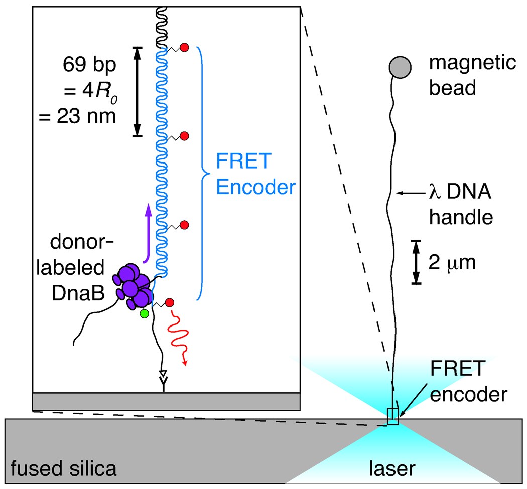
Figure 2.
Illustration of the experiment, approximately to scale. The FRET encoder is tethered via an anti-digoxigenin/digoxigenin linkage to a fused-silica cover slip. A biotinylated λ DNA handle, ligated to the untethered end of the encoder, is attached to a streptavidin-coated magnetic bead. A 0.5–3.0 pN vertical magnetic force is applied to the bead, pulling it away from the surface and aligning the encoder with the optical axis. Donor-labeled DnaB helicase diffuses into the focus and loads onto the free 5′ tail of the encoder. As the encoder is unwound, the moving laser-excited donor passes one acceptor after another, inducing long-wavelength fluorescence via FRET. The resulting periodic acceptor signal reports on the motion. A spacing of 4R0 between acceptor dyes was chosen so that the donor would be within 2R0 of only a single acceptor at any given time.

