
Figure 1.
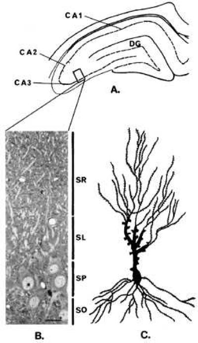
Localization of the hippocampal stratum lucidum area used for electron microscopic studies. (A) Durcupan-embedded blocks containing 100-μm-thick sections of the dorsal hippocampus were trimmed at the level of the CA3 region (see box). (B) Semithin sections (1.5 μm) stained with toluidine blue were used as a guide to further trim the blocks to an area containing the stratum lucidum (SL). (Bar = 17.5 μm.) (C) Camera lucida tracing of a representative pyramidal neuron impregnated with the “single” section Golgi method (13). DG, dentate gyrus, SO, stratum oriens, SP, stratum pyramidale, SL, stratum lucidum, SR stratum radiatum.