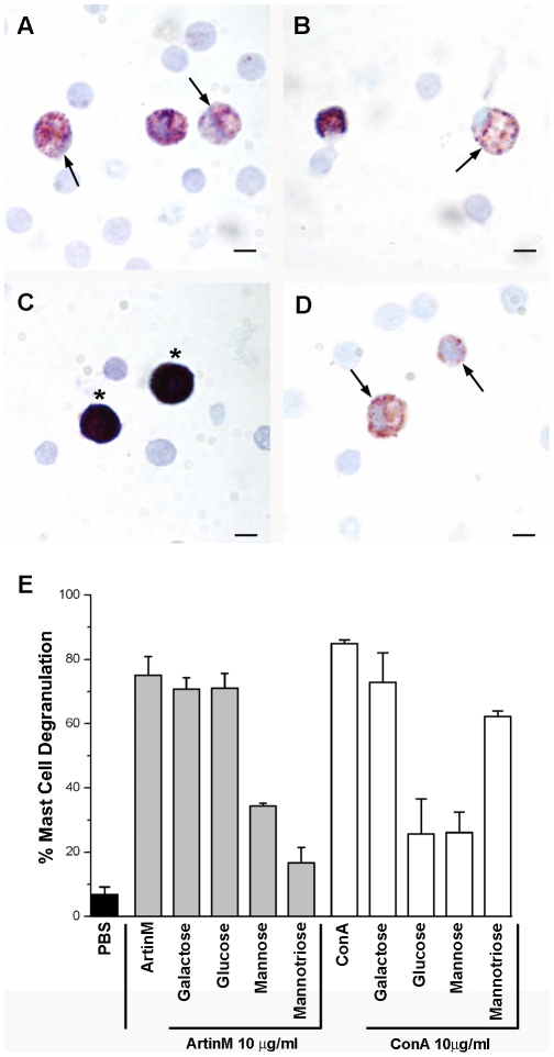
Figure 1. ArtinM and ConA degranulate peritoneal mast cells in vitro in a lectin specific manner.
The total cell population from the peritoneal lavage was incubated with 10 µg of ArtinM (A) or ConA (B). Control cells were incubated in PBS (C). Compound 48/80 (D) was used as a positive control for degranulation. Stained with Toluidine blue. (Arrows, degranulated mast cells; Asterisks, intact mast cells) Bars = 10 µm. Quantification of peritoneal mast cell degranulation (E). The data shown is the average±SD of 3 separate experiments.

