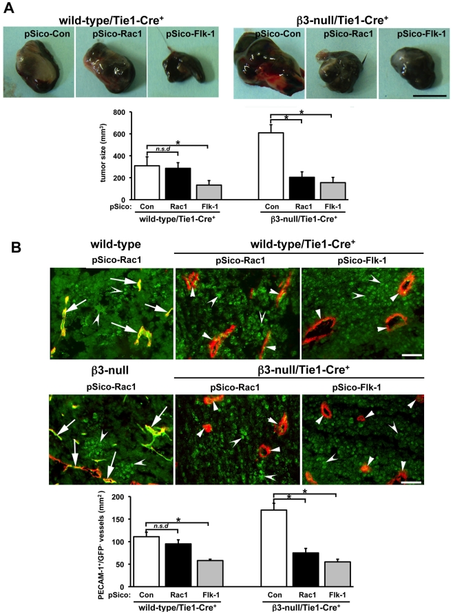
Figure 3. Endothelial-specific Rac1-depletion does not impair tumor growth wild-type mice but does in β3-null mice.
A. Murine B16F0 cells (106) were injected subcutaneously into the flanks of wild-type/Tie1-Cre+ or β3-null/Tie1-Cre+ mice. Lentiviral vector suspensions (106 i.u/ml) of pSico-Con, pSico-Rac1 and pSico-Flk-1 were injected intratumorally on days 5 and 10 after tumor cell injection. Representative macroscopic appearance of 14-day-old B16F0 pSico-Con-, pSico-Rac1- and pSico-Flk-1- treated melanomas in both genotypes. Scale bar: 5 mm. Bar graph shows mean tumor volume per mm3 (+ s.e.m.). Tumor size was reduced significantly in pSico-Flk-1-treated mice of both genotypes (*P<0.05) and in pSico-Rac1 treated β3-null/Tie1-Cre+ but not in pSico-Rac1-treated wild-type/Tie1-Cre+ mice (n.s.d, no significant differences). N = 4–6 animals per condition. B. Representative merged images of PECAM-1 (red) and GFP (green) -immunostained sections from pSico-Rac1-treated B16F0 tumors grown in wild-type, wild-type/Tie1-Cre+, β3-null and β3-null/Tie1-Cre+ mice. PECAM-1-positive staining identified endothelium. GFP-positive staining was observed in tumor cells (concave arrowheads) and in PECAM+ endothelium (arrows) of blood vessels in B16F0 tumors from wild-type and β3-null control mice, indicating successful pSico-Rac1 lentivirus infection in vivo. Loss of GFP detection in most PECAM-1-positive microvessels (small arrowheads), but not in B16F0 tumor cells, was observed in pSico-treated tumors grown in wild-type/Tie1-Cre+ and β3-null/Tie1-Cre+ mice, indicating successful endothelial-specific Cre recombination in vivo. Scale bar: 10 µm. Bar graph shows mean numbers of PECAM-1+/GFP− vessels per unit area of tumor section per mm2 (+ s.e.m). Blood vessel density was reduced significantly in pSico-Flk-1-treated mice of both wild-type/Tie1-Cre+ and β3-null/Tie1-Cre+ mice (*P<0.05) and in pSico-Rac1-treated β3-null/Tie1-Cre+ mice but not pSico-Rac1-treated wild-type/Tie1-Cre+ mice (n.s.d, no significant differences). N = 4 animals per condition.

