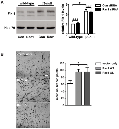
Figure 9. Endothelial Rac1-depletion does not affect the expression of Flk-1.
A. Western blot analysis of Flk-1 expression levels in primary wild-type and β3-null endothelial cells transfected with scrambled (Con siRNA) or Rac1 (Rac1 siRNA) siRNAs. Although Flk-1 expression was increased significantly in β3-null endothelial cells when compared with wild-types (*P<0.01), Rac1-depletion did not affect Flk-1 levels (n.s.d) in either genotype. Bar graph represents mean (+ s.e.m.) of Flk-1 expression relative to Hsc-70 loading control (N = 3–4 independent experiments). B. HUVEC were transfected with vector only, a wild-type Rac1 construct (Rac1 WT), or a constitutively active Rac1 construct (Rac1 QL) and seeded on confluent fibroblasts. Tubules were visualized by PECAM1 staining 5 days after seeding. The mean number of branch points (+SEM) is shown in the accompanying bar chart. (n = 12 microscopic fields per condition). Scale bar: 100 µm.

