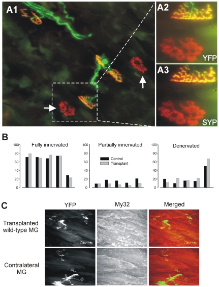
Figure 3. B6.SOD1 motor neurons fail to completely reinnervate wild-type muscles.
A1. Low magnification view of wild-type muscle transplanted into an B6.SOD1 animal 2 months earlier. The host wildtype animal expressed YFP in neuronal membranes (green) while ACHRs are labeled with α−Btx (red). Denervated endplates are indicated by white arrows. A2–3. Higher magnification and rotated view of area indicated in A1 by broken lines. Panels illustrate complete occupation of upper endplate by nerve terminal (A2, YFP) and synaptophysin staining (A3, SYP) and complete denervation of lower endplate. B. Percentages of fully and partially innervated endplates and denervated endplates for transplanted and contralateral control MG muscles. Bar pairs correspond to the same animals. C. Low magnification views of YFP-labeled motor terminals and My32–stained muscle fibers show that fast muscle fibers regenerated in wild-type MG muscle transplanted into B6.SOD1 animals and showed similar levels of denervation.

