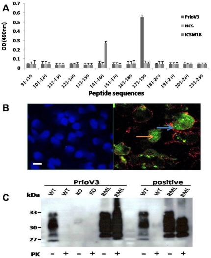
Figure 1. PrioV3 binds to both native and denatured prion proteins.
(A) PrioV3 antibody was screened with 20-mer amino acid sequences spanning the 91–230 region of the prion protein and was shown to bind a region between 170 and 19. ICSM18 that binds to a region between 141 and 160 was used as positive control and normal camel serum (NCS) was used as negative control. Goat anti-llama IgG-HRP was used as secondary detection antibody. Anti-PrP responses were measured in peptide ELISA. Error bars represent the mean antibody level derived from n = 8 wells. (B) PrioV3 antibody bound to native PrPC inside the cytoplasm of N2a cells (orange arrow), in contrast with ICSM35 that strictly stained cell membrane-bound PrPC (blue arrow). Florescence microscopy was performed and images from each source [FITC (450–490 nm), Texas red (510–560 nm) and DAPI (330–380 nm)] were collected. As control, N2a cells were stained with the secondary anti-llama IgG (green) and anti-mouse IgG antibody (red), omitting the primary antibodies. Scale bar = 25 µm (C) PrP wild type, knock-out and RML-infected brain homogenate were digested with PK and tested for reactivity PrioV3. PrioV3 strongly bound both PrPC and PrPSc in brain homogenates (pre- and post-PK). For comparison, ICSM35, raised in mouse (positive), was incubated with brain homogenates prepared from RML-infected and normal brain tissue with and with no PK digestion.

