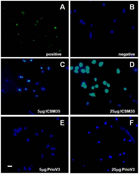
Figure 7. ICSM35 but not PrioV3 anti-prion antibody leads to neurotoxicity N2a cells following antibody treatment.
N2a cells were assessed for neurotoxic effects by immunofluorescence imaging following treatment with PrioV3 or ICSM35. (A) Proprietary positive TUNEL stain and (B) antibody-free tissue culture medium were used as controls. Following treatment with 5 and 25 µg PrioV3 or ICSM35, TUNEL staining was only observed when cells were treated with ICSM35 (C & D). PrioV3 did not elicit DNA fragmentation and toxic effects were not observed (E & F). Cell nuclei were also stained with DAPI. Scale bar = 5 µm. Representative of three experiments.

