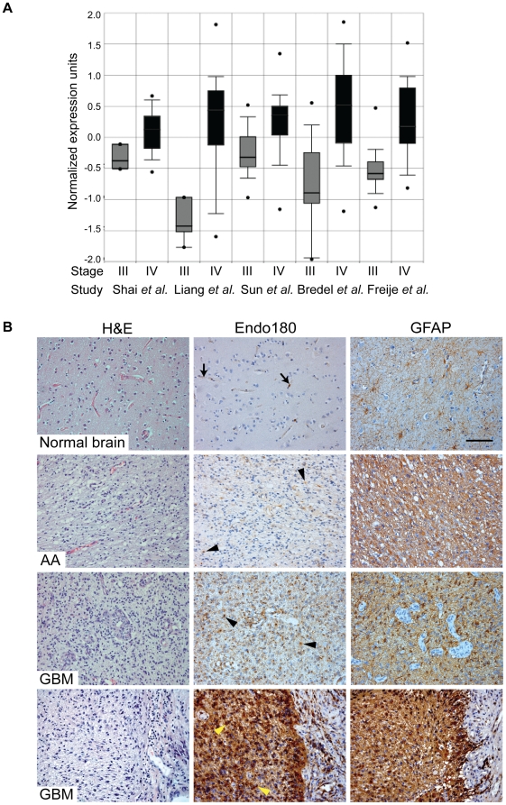
Figure 1. Endo180 expression is highly upregulated in GBMs.
(A) Normalized expression of Endo180 in grade III gliomas (astrocytomas, oligodendrogliomas and oligoastrocytomas) and grade IV gliomas (GBMs). Box plots were created by ONCOMINE™ from five independent expression array studies. p-values were 5.6×10⁻⁵ (Shai et al.) [22], 1×10⁻⁶ (Liang et al.) [21], 2.2×10⁻¹² (Sun et al.) [23], 5.9×10⁻⁸ (Bredel et al.) [19] and 2.3×10⁻¹² (Freije et al.) [20]. (B) FFPE whole tissue sections of normal brain (2 samples) and grade III (2 samples) or grade IV gliomas (9 samples) were H&E stained or immunostained for Endo180 (mAb 39.10) and glial fibrillary acidic protein (GFAP). Representative images of the temporal lobe of normal brain showing weak expression of Endo180 in some cells associated with the vasculature (arrows); a grade III anaplastic astrocytoma (AA) showing weak Endo180 expression in GFAP-positive tumor cells (arrowheads), two grade IV glioblastomas (GBM) showing strong Endo180 expression in tumor cells (black and yellow arrowheads). Scale bar, 100 µm.

