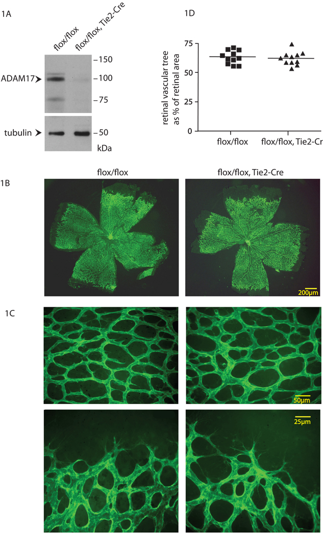
Figure 1. Evaluation of developmental retinal angiogenesis in Adam17flox/flox/Tie2-Cre mice.
(A) Western blot analysis shows a strong reduction of ADAM17 protein levels in primary endothelial cells isolated from an Adam17flox/flox/Tie2-Cre mouse compared to an Adam17flox/flox littermate control, with tubulin serving as a loading control (see materials and methods). (B,C) Whole-mount isolectin B4 staining of the developing retinal vasculature of 6 day-old Adam17flox/flox/Tie2-Cre and Adam17flox/flox littermates. Panel C shows higher magnification photomicrographs of the density of the vascular web (top panels) and the leading edge of the vascular tree (lower panels). (D) The ratio of the surface area of the developing vascular tree over that of the retina showed no significant difference (WMW test, p=0.76) in the extent of developmental retinal angiogenesis in Adam17flox/flox/Tie2-Cre mice (n=11) compared to Adam17flox/flox controls (n=11).

