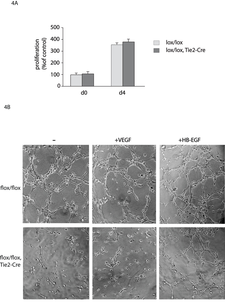
Figure 4. Endothelial cell proliferation and tube formation assays.
(A) Proliferation assay and (B) tube formation assay with endothelial cells isolated from Adam17flox/flox/Tie2-Cre mice or Adam17flox/flox controls (see materials and methods). There was no significant difference in proliferation at day 4 (d4) of the culture plated a comparable density on day 0 (d0) (A), but a clear decrease in tube formation was seen in endothelial cells from Adam17flox/flox/Tie2-Cre mice compared to controls (B), which could be largely rescued by treatment with 5ng/ml HB-EGF, but not with 5ng/ml VEGF-A.

