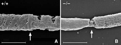
FIG. 3.
Cold-field emission scanning electron micrographs of demembranated sperm tails focusing on the principal piece-midpiece junction. A) shows the annulus (arrow) of a WT sperm, while B shows the gap (arrow) created by the missing annulus of a Sept4−/− sperm. In both pictures, the mitochondrial sheath of the midpiece is to the left of the arrow, and the fibrous sheath of the principal piece is to the right of the arrow. Bar = 1.0 μm.

