
Figure 1.
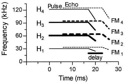
Schematized sonagram of the mustached bat orientation pulse (solid lines) and the Doppler-shifted, time-delayed echo (dashed lines). The four harmonics (H1–H4) of both pulse and echo each contain a long constant frequency component and a short FM component (FM1–4). The thickness of the lines indicates differences in relative amplitude of harmonics. According to their heteroharmonic combination-sensitivity for echolocation pulse–echo pairs, three major types of FM1–FMn neurons can be distinguished: FM1–FM2, FM1–FM3, and FM1–FM4 (redrawn from ref. 15).