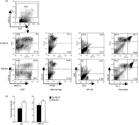
Figure 5.
Cell recruitment and neutrophil chemokine production in lung tissue of BALB/c and C57BL/6 mice after infection with Chlamydia muridarum. Groups of three mice were inoculated intranasally with 8000 inclusion-forming units (IFU) of C. muridarum and day 2 post-infection lungs were harvested, single cell suspensions were created and cell types were enumerated by fluorescence-activated cell sorter analysis. Two independent experiments were carried out. Dot-plot from one representative mouse shows double staining of surface markers for lung inflammatory cells (a). Chemokine secretion in lung tissue of BALB/c mice and C57BL/6 mice at day 2 after infection with C. muridarum. Lung homogenates were the same samples prepared for Fig. 3. The chemokines in lung homogenates were analysed by enzyme-linked immunosorbent assay. The data represent the mean ± SE from eight individual mice. One of three independent trials with similar results is shown. *P < 0·05 (b).

