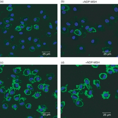
Figure 5.
Confocal microscopy of [Nle4, DPhe7]-α-melanocyte-stimulating hormone (NDP-MSH)-treated and untreated mature and immature dendritic cells (DCs). The DCs generated from monocytes after 6 days in culture in RPMI-1640 containing granulocyte–macrophage colony-stimulating factor and interleukin-4 were stained with fluorescein isothiocyanate-conjugated major histocompatibility complex (MHC) class I (HLA-A,B,C). Low numbers of immature DCs stained positive, with green surface-staining of cell membranes (a and b). After treatment with lipopolysaccharide, MHC class I was up-regulated and dendritic processes were exhibited on cell surfaces (c and d). No significant difference in the cytological appearance was observed between NDP-MSH-treated (b and d) or untreated (a and c) cells. The horizontal bar indicates 25 μm.

