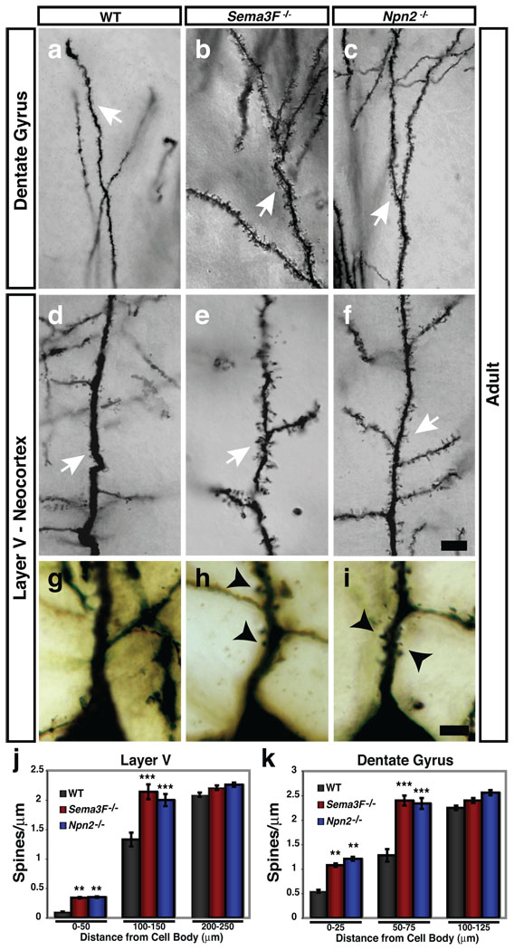
Figure 1. Sema3F and Npn-2 regulate dendritic spine number, distribution and morphology in adult layer V pyramidal neurons and dentate gyrus granule cells in vivo.
a–f, Golgi stained Sema3F−/− (b, e) and Npn-2−/− (c, f) brains show that DG GC and layer V pyramidal apical dendritic spines (white arrows) are more numerous as compared to WT (a, d). g–i, Layer V pyramidal neurons have more spines on primary apical dendrites 0–25 µm from the soma in Sema3F−/− (h) and Npn-2−/− (i) mutants as compared to WT mice (g). j, k, Quantification of spine density 0–50µm from the cell body on layer V pyramidal primary apical dendrites (WT, 0.10 ±0.01; Sema3F−/−, 0.34 ±0.01; Npn-2−/−, 0.35 ±0.01 spines/µm) and 0–25µm from the cell body on DG GC primary dendrites (WT, 0.55 ±0.03; Sema3F−/−, 1.08 ±0.04 and Npn-2−/−, 1.21 ±0.04 spines/µm). There is a significant increase in spine number on dendritic segments located 100–150µm from the cell body in layer V (WT, 1.33 ±0.14; Sema3F−/−, 2.14 ±0.13; Npn-2−/−, 2.00 ±0.11 spines/µm) and 50–75µm from the DG neuron cell body (WT, 1.28 ±0.13; Sema3F−/−, 2.40 ±0.11; Npn-2−/−, 2.34 ±0.12 spines/µm) in these mutants. There is no significant difference in spine density at 200–250µm or 100–125µm from the cell body in layer V and DG neurons, respectively. Error bars, ±SEM, ANOVA, post-hoc Tukey in j and k; **, p=0.01; ***, p=0.001 compared to WT. Scale bars: 10 µm in f for a–f and 2.5 µm in i for g–i.

