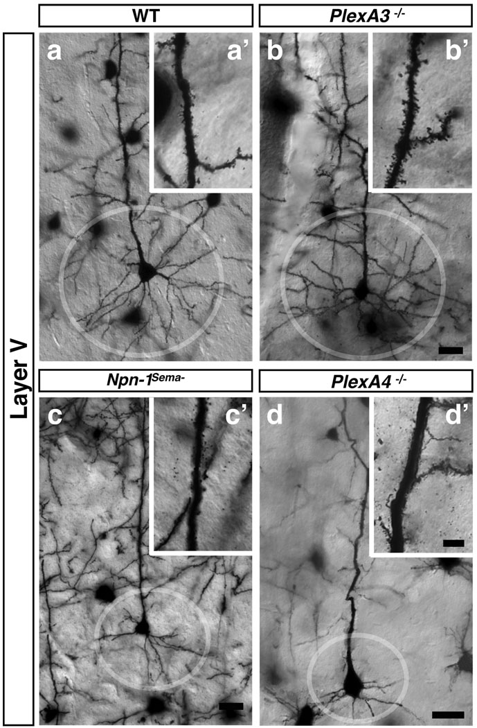
Figure 4. Distinct Sema3–Npn/PlexA signaling modules regulate apical dendrite spine morphology and basal dendrite process complexity.
a–d, Golgi-labeled adult brains illuminate basal dendritic morphologies in cortical layer V pyramidal neurons from WT (a, circle), PlexA3−/− (b), Npn-1Sema− (c), and PlexA4−/− (d) mice. a’–d’, show spine morphologies from neurons in a–d. Scale bars: 10 µm in d for a–d and 4 µm in d’ for a’–d’.

