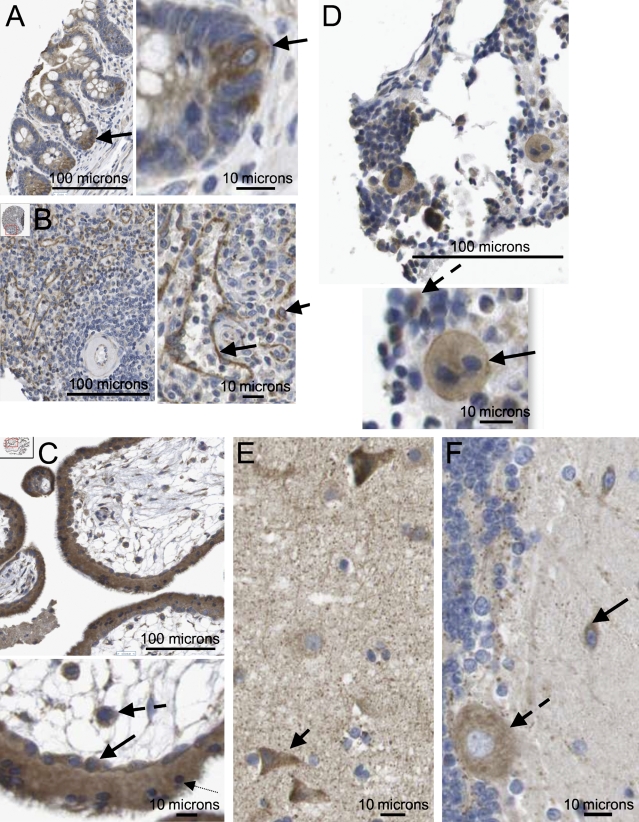
Figure 2.
Axotrophin/MARCH-7 protein expression in selected tissue types. (A) Small intestine showing strong positive, precise, and consistent cytoplasmic staining of crypt basal progenitor cells (arrow); nucleus is negative. Adjacent cells higher up the crypt show relatively weak cytoplasmic staining. (B) In the spleen, the sinusoidal endothelium shows strong positive, precise, and consistent staining of the cytoplasm (longer arrow). In the splenic pulp, most lymphocytes are negative, apart from a small number of cells with specific cytoplasmic staining (shorter arrow). Small arteriole is negative. (C) First trimester placenta; here, there is strong, precise, and consistent positive staining of both the cytotrophoblast (solid arrow) and the syncitiotrophoblast (dotted arrow); a few cells also show nuclear positivity. Cells within the vascular mesoderm of the villus are also strongly positive (dashed arrow). (D) In bone marrow, megakaryocytes are present and show clear moderate granular expression of axotrophin/MARCH-7, which is increasing at the plasma membrane (solid arrow). Some 40% of small mononuclear cells also show cytoplasmic positivity (dashed arrow). (E) In the CNS, the cerebral cortex neurons show a range of moderate to strong cytoplasmic positivity (arrow). A fine network of lightly stained neuropils is evident. The glia is negative. (F) CNS cerebellum showing moderate cytoplasmic positivity in neurons (solid arrow) and Purkinje cells (dashed arrow). The glia is negative.

