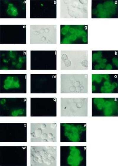
Figure 2.

Immunofluorescent microscopic indirect labeling experiments with transfected DAMI cells, by using the several primary anti-peptide antibodies described in Materials and Methods and directed toward the epitopes shown in Fig. 1. [The N2 and N3 antibodies did not exhibit unique specificity for their epitope (21), unlike the other antibodies, and were therefore not included]. (a–d) DAMI cells transfected with PS-2 and immunolabeled with antibody N1: (a) live cells; (b) live cells labeled in the presence of excess specific soluble oligopeptide conjugate for N1; (c) the Nomarski image of b; (d) fixed and permeabilized cells. (e–g) DAMI cells transfected with PS-1 and immunolabeled with antibody I1: (e) live cells; (f) the Nomarski image of e; (g) fixed and permeabilized cells. (h–k) Same as a–d except that immunolabeling was of PS-1-transfected cells with antibody L3 and in i the specific soluble oligopeptide conjugate for L3 was used for inhibition. (l–o) DAMI cells transfected with PS-2 and immunolabeled with antibody L2: (l) live cells; (m) live cells labeled in the presence of excess specific soluble oligopeptide conjugate for L2; (n) the Nomarski image of m; (o) fixed and permeabilized cells. (p–s) DAMI cells transfected with PS-1 and immunolabeled with antibody L1: (p) live cells; (q) live cells labeled in the presence of excess specific soluble oligopeptide conjugate for L1; (r) the Nomarski image of q; (s) fixed and permeabilized cells. (t–v) DAMI cells transfected with PS-1 and immunolabeled with antibody C1: (t) live cells; (u) the Nomarski image of t; (v) fixed and permeabilized cells. (w–y) Same as t–v except with cells transfected with PS-2.
