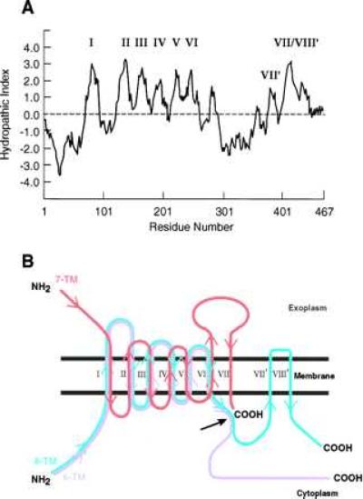
Figure 3.

(A) The Kyte-Doolittle hydropathy plot for PS-1 (1) by using a window of 15 residues. The roman numerals indicate the hydrophobic sequences serving as TM spanning stretches in B (see text). (B) The topographies of the three proposed models of PS proteins, the 6- (magenta), 7- (red), and 8- (blue) TM spanning models. The correspondingly colored arrows indicate the polypeptide chain directions starting from the NH2 terminus (NH2) toward the COOH terminus (COOH) for each model. The long black arrow indicates the region of the proposed endoproteolytic cleavage of the PS proteins (see text).
