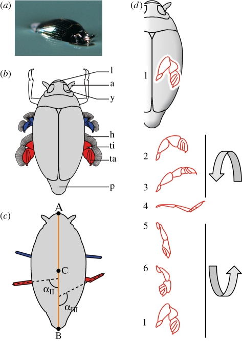
Figure 1.
(a) Close-up picture of a whirligig beetle (Gyrinus substriautus) at rest on the water surface. (b) Schematic drawing of a whirligig beetle in dorsal view. Middle legs are represented in blue, and hind legs in red. Forelegs that remain folded under the body during swimming are shown in white. a, Antennae; h, hairs of the swimming blades; l, labium; p, pygidium; ta, tarsi; ti, tibia; y, eye. (c) Whirligig beetle with (A) the end of the labium, (B) the end of the pygidium and (C) the centre of the body. The orange line represents the body axis. Middle legs are represented in blue, and hind legs in red. The angles αII and αIII represent the maximum angles of the hind legs measured for leg kinematics types II and III, respectively (see the text for more detailed explanation). (d) Kinematics of the right hind leg (in red) during a stroke, seen from above (modified from Nachtigall 1961). Each position of the leg is separated by 2 ms. In (4), the position of tarsi is that produced by type III leg kinematics; for type II leg kinematics, the tibia and tarsi stay in the same axis. Three-dimensional arrows indicate the rotation of the leg during the stroke.

