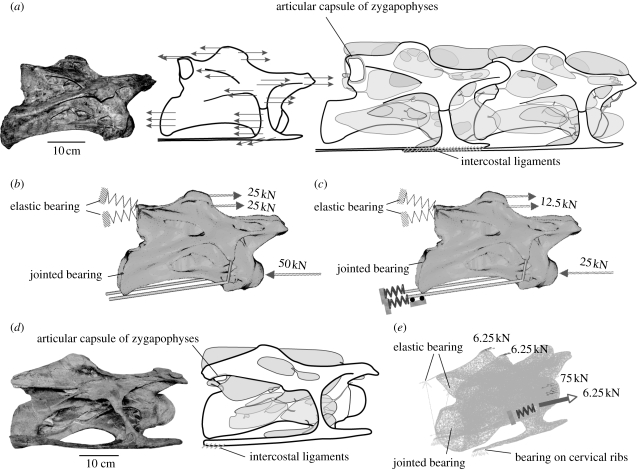
Figure 1.
Used soft-tissue insertions (after Schwarz et al. 2007; Schwarz & Frey 2008) and simplified models for FE analysis. (a) Fourth cervical vertebra of Brachiosaurus brancai (MB.R.2080.25), line drawing with vectors of assumed axial cervical muscles indicated in their insertion sites, and reconstruction of fourth and fifth cervical vertebrae of B. brancai with pneumatic diverticula and articular soft tissues; right lateral view. (b) Model for the fourth cervical vertebra of B. brancai used in the FEA with applied forces during extension. (c) Same model with applied forces during ventral flexion and suspension on the cervical ribs. (d) Mid-cervical vertebra of Diplodocus sp. (SMA L25-3) in right lateral view and with reconstructed pneumatic diverticula and soft tissues used for FEA in the load case of lateral flexion. (e) Model for the mid-cervical vertebra of Diplodocus used in the FEA with applied forces during lateral flexion.

