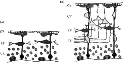
Figure 1.
Development of the cerebral cortex in normal mice. (a) The pre-plate stage. CR, Cajal–Retzius cells lying beneath the pia; SP, subplate cells; VZ, ventricular zone containing germinal neurons and glial cells. The glial fibres extend from the VZ to the outer margin of the pre-plate. (b) The cortical plate (CP) stage. Post-mitotic neurons leave the VZ and crawl along glial fibres, past the intermediate zone (IZ) of the cortex and the SP to form the CP between the SP and the marginal zone (MZ) containing the CR cells. Adapted from Rice & Curran (1999) with the permission of the authors.

