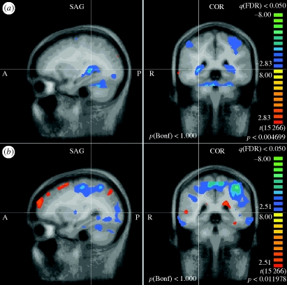
Figure 4.
Hippocampus/caudate. (a) PELFMF post- to pre-condition. This image shows a significant decrease in activity within the hippocampus/caudate area following PELFMF exposure when compared with pre-exposure. Due to the relatively poor spatial resolution, it is difficult to say exactly from which structure(s) this activity is originating. (b) Sham post- to pre-condition. No difference is seen due to time alone in the sham condition.

