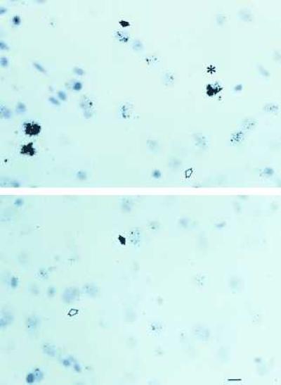
Figure 2.

Photomicrographs of sections through the VMN of ovariectomized rats treated with E for 18 hr (Upper) or control ovariectomized female rats (Lower) hybridized with 3H-labeled antisense probe I-A. The open arrowheads point to unlabeled cells, the filled arrows point to labeled cells, and the asterisk indicates a highly labeled cell containing a focus of silver grains. (Bar = 20 μ.)
