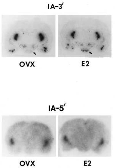
Figure 4.

Representative film autoradiographs of brain sections of an OVX and 18 hr E-treated female rat hybridized with probe I-A3′ and I-A5′-labeled with 32P. Note that in the VMN (identified by arrow) of the E2-treated animal, a hybridization signal is detectable with probe I-A of similar intensity as in the reticular thalamic nucleus, indicating the appearance of PPEIA-3′ RNA in the VMN following E treatment. Exposure time: Upper; 1 day; Lower, 5 days. The Lower panel was exposed for a longer period to show that probe I-A5′ does label the striatum as expected.
