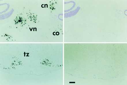
Figure 5.

Low power photomicrographs showing the hybridization of probe I-A in the brainstem. The left panels show hybridization with the antisense probe, the right panels show hybridization with the sense strand probe. cn, Cerebellar nuclei; vn, vestibular nuclei; co, cochlear nucleus; tz, trapezoid body. (Bar = 200 μ.)
