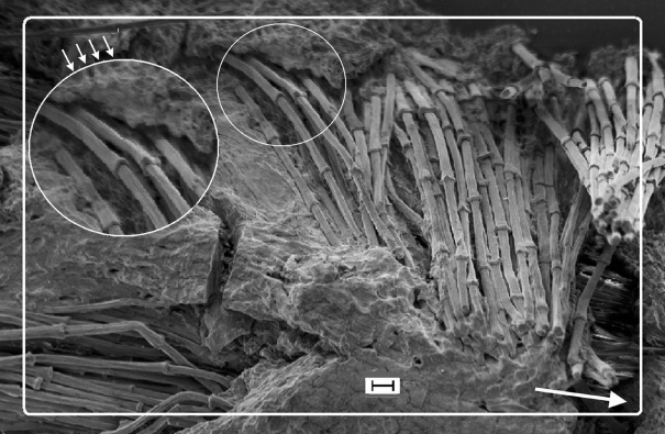
Figure 3.
SEM of biodegraded feather rachis of Gallus gallus (resin embedded and etched). Circumferential fibres (syncitial barbules), identical to the longitudinal fibres (see below in the figure), wound round the outer ‘layers’ of the rachidial cortex. The matrix is partially degraded and shows the honeycomb-like structure in which the fibres are embedded in life (small arrows). Detail shows fibres, comparable to steel rebars in concrete (see text). Arrow shows long axis of rachis. Scale bar, 10 µm.

