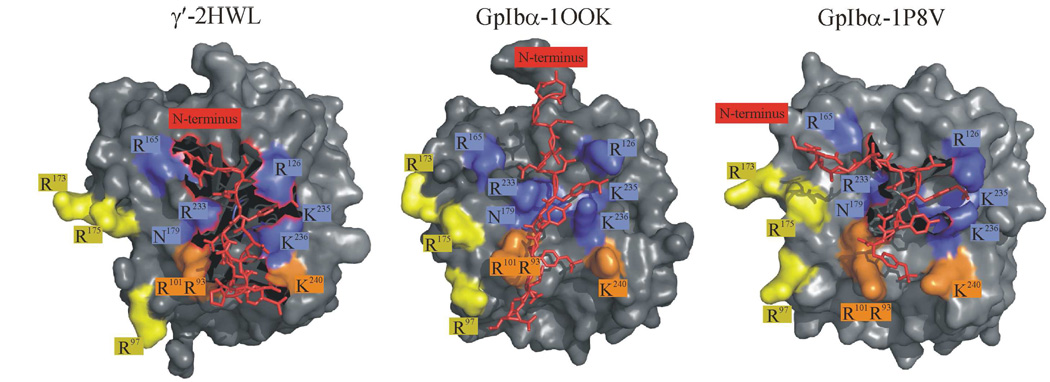
Figure 12.
Crystallographic views of the γ′ peptide (2HWL) and GpIbα (1OOK and 1P8V) bound to thrombin’s ABE-II. A prominent feature involves GpIbα YP 276 and γ′ YP 418 engulfed by a electropositive pocket formed by IIa R126, K235, and K236. In addition, GpIbα D277 and γ′ D419 reside near IIa N179. Finally, YP 279 and γ′ YP 422 point toward IIa K236. These structures illustrate similar bound characteristics for the central regions of the bound peptide, however differ in regard to the termini. These figures were created using PyMol (11).

