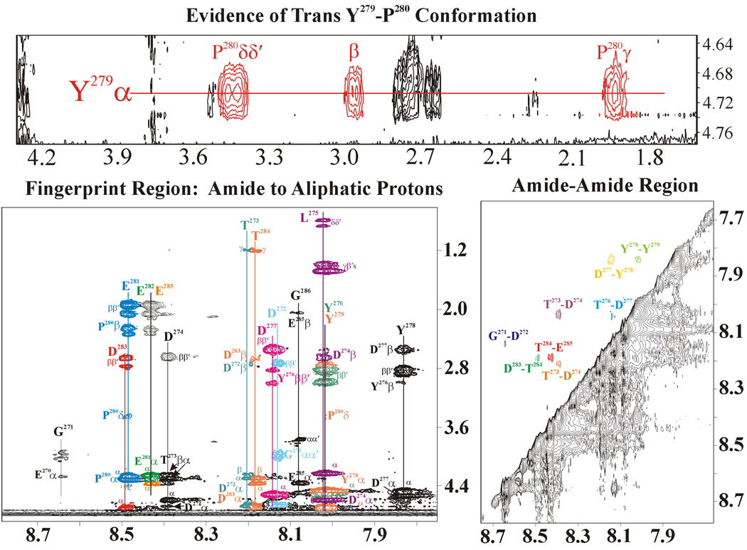
Figure 4.
2D trNOESY spectrum of the unphosphorylated GpIbα (1.5 mM) bound to IIa (0.150 mM). The top panel illustrates the evidence for a trans Y279-P280 peptide bond. The bottom panels demonstrate that GpIbα exists in an extended structure when bound to IIa due to the presence of only nearest neighbor NOEs. The NOEs are color coded according to the corresponding residue.

