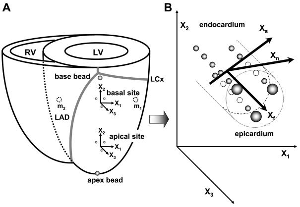
Fig. 1.
A: schematic representation of the heart. LV, left ventricle; RV, right ventricle; X1, circumferential axis; X2, longitudinal axis; X3, radial axis; LAD, left anterior descending coronary artery; LCx, left circumflex coronary artery; and m1, m2: intramyocardial markers implanted at the level of the basal bead set in the LV lateral wall (m1) and the septum (m2). B: schematic representation of local fiber-sheet axes. Xf, fiber axis; Xs, sheet axis; and Xn, axis oriented normal to the sheet plane. The Xf, Xs, and Xn axes present a Cartesian system.

