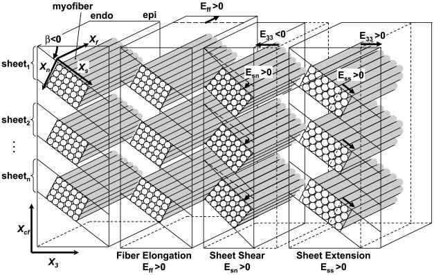
Fig. 2.
Schematic representation of fiber-sheet remodeling strains. Each cylinder represents a myofiber. Myofibers are organized into laminar “sheets,” which are approximately four cells thick and roughly stacked from apex to base (15). Sheet angle (β) is measured with reference to the positive X3, with a positive angle defined as rotation toward the positive cross-fiber axis (Xcf). Therefore, the sheet angle is negative in this diagram (β < 0). Positive Eff, Esn, and Ess represent fiber elongation, sheet shear, and sheet extension, respectively. Because the sheet angle is negative (β < 0), a positive sheet shear (Esn > 0) is associated with reduction in radial remodeling strain or wall thinning (E33 < 0). Sheet extension (Ess > 0) may be accounted for by myofiber rearrangement within the sheet plane, or interdigitation, in addition to an increase in myocyte diameter (7), and is associated with increase in radial remodeling strain, or wall thickening (E33 > 0). Endo, endocardium; epi, epicardium.

